WELCOME TO THIS GREAT AND EXCITING EDUCATIONAL BLOG

Welcome to my TEAS Exam Prep blog. I hope it will help you prepare very well for the pre-nursing entrance exam, popularly known as the TEAS exam. This is one of the entrance exams required by some schools for those who want to pursue careers in the nursing field in the USA. I will attempt to break down the review materials into manageable parts so that you can systematically and efficiently prepare for the test with less stress. I will guide you to prepare for the entire content of the test. Hopefully, you will be able to pass after going through these series.


Best of luck!


To support this blog and my effort to make more quality free educational videos you can make a donation by clicking on the link below. Together, we can change somebody's life one person at a time around the globe.
Become a Patron!

Tuesday, May 27, 2014

GAS LAWS

The following are the basic laws that govern the behavior of gases.
1. Boyle's law: This states that at a constant temperature, the pressure of a gas is inversely proportional to its volume. This means that as the pressure increases, the volume decreases and vice versa.

(Initial Pressure) x (Initial Volume) = (Final Pressure) x (Final Volume)

Examples: 

  • As the pressure in a balloon is increased, it expands continuously until it can no longer do so and then burst open. 
  • Air rushes into the lungs from outside when the chest cavity expands to increase its volume because the pressure within the thoracic cavity decreases. When the volume of the thoracic cavity decreases, the intra-thoracic pressure increases and the air is pushed out. 
2. Charles' law: This states that at a constant pressure, the volume of a gas varies directly as its temperature. This means that when the temperature increases, the pressure also increases and vice versa.

   Initial Volume      =   Final Volume      
Initial Temperature       Final Temperature

Examples: 
If you increase the temperature on a balloon at atmospheric pressure, the volume increases as well.

3. Gay-Lussac's law: This states that at a constant volume, the pressure of a gas varies directly as its temperature. This means that when the temperature increases, the pressure also increases and vice versa.

   Initial Pressure      =   Final Pressure      
Initial Temperature       Final Temperature

Monday, May 26, 2014

TEMPERATURE CONVERSIONS

Here are the formulas for converting from one temperature unit to another.
C is Celsius
F is Fahrenheit
K is Kelvin 
1.   C = 5/9 (F-32) or C = (F-32)/1.8
2.    F = 9/5 (C+32)  or  F = (C+32)*1.8
3.    K = C + 273
Note that the Kelvin temperature has no negative values. 

PHASE DIAGRAMS AND PHASE CHANGES

What is a phase diagram?
Phase diagrams show the preferred physical states of matter at different temperatures and pressure. Within each phase, the material is uniform with respect to its chemical composition and physical state. 


Click on this for "Practice exercise on phase diagram"
Click here for the Answers to practice exercise

PHASE CHANGES
This is a link to a website that has a good explanation on phase changes of water. I recommend that you read it. It will be good to memorize and understand what happens in the various regions. Note that when there is a change of state, the temperature remains unchanged for sometime.

Diagram of phase changes

Friday, May 23, 2014

QUESTIONS FOR THE DAY

  1. Calculate the amount of heat needed to increase the temperature of 250g of water from 20C to 56C. Specific heat capacity = 4.18 J oC-1 g-1
  2. Calculate the specific heat capacity of copper given that 204.75 J of energy raises the temperature of 15 grams of copper from 25o C to 60C.
  3. 216 J of energy is required to raise the temperature of aluminium from 15o to 35oC. Calculate the mass of aluminium. (Specific Heat Capacity of aluminium is 0.90 JoC-1g-1).
  4. The initial temperature of 150g of ethanol was 22oC. What will be the final temperature of the ethanol if 3240 J was needed to raise the temperature of the ethanol?
    (Specific heat capacity of ethanol is 2.44 JoC-1g-1).
Answers:
  1. q = 250 x 4.18 x (56 - 20) = 37,620 J or approx. 38 kJ
  2. 204.75 = 15 x C x (60 - 25)
    C = 0.39 JoC-1 g-1
  3. 216 = m x 0.90 x (35 - 15)
    m = 216 ÷ 18 = 12g
  4. 3240 = 150 x 2.44 x (Tf - 22)
    Tf = 30.9oC

QUANTITY OF HEAT

The quantity of heat energy (Q) gained or lost by a substance is equal to the mass of the substance (m) multiplied by its specific heat capacity (C) multiplied by the change in temperature (final temperature - initial temperature)


Q = m x C x (Tf - Ti)

Memorize this formula well.

  • Specific Heat Capacity (C) of a substance is the amount of heat required to raise the temperature of 1 gram of the substance by 1C (or by 1 K).
  • Heat capacity (H) is defined as the mass multiplied by the specific heat capacity.        H = m x C
  • The quantity of heat can therefore be defined as heat capacity multiplied by the change in temperature. Q = H x C
Example:
Calculate the quantity of heat needed to raise the temperature of 250 grams of water from 20C to 56C. (Heat capacity of water is Cg = 4.18 J oC-1 g-1)

Solution: Q = m x C x (Tf - Ti) 
m = 250 g; Tf = 56oC; Ti = 20oC

q = 250 x 4.18 x (56 - 20) 
q = 250 x 4.18 x 36 
q = 37620 J = 38 kJ

MODES OF HEAT TRANSFER

Heat is a form of energy which transfers between bodies which are under thermal interactions. Heat transfer occurs when a temperature difference occurs between two bodies or a body with its surroundings. 
There are three modes of heat transfer:
  1. Conduction
  2. Convection and
  3. Radiation
CONDUCTION: Conduction is the mode of heat transfer which occurs from one part of a substance to another part within the substance itself or with another substance which is placed in physical contact. 
CONVECTION: Heat transfer occurs within a fluid itself and it is carried out by transfer of one fraction of the fluid to the remaining portion. 
RADIATION: This mode of heat transfer does not require any medium to occur. Every matter with a temperature above absolute zero will emit energy in the form of electromagnetic waves called radiation.

Friday, May 16, 2014

TYPES OF TISSUES

A tissue is a group of cells designed to perform a particular function. There are four types of tissues.

1. Epithelium (Epithelial tissue) - These are cells that line surfaces of biological structures, such as lining of the intestine or the skin covering.

There are three types of epithelial tissues classified based on their shapes:
(a) Squamous epithelium - flat cells
(b) Cuboidal epithelium - Look like cubes
(c) Columnar epithelium - Look like columns or pillars

They can also be classified based on the number of layers:
(a) Simple epithelium: Only one layer of cells
(b) Stratified epithelium: Many or multiple layers of cells
(c) Pseudoepithelium: This epithelium has only one layer but appears under the microscope as multiple layers
(d) Transitional epithelium -This epithelium has many layers of cells with various shapes. It lines the ureters and bladder and functions in the distension of these structures.

Some of the cuboidal/columnar epithelial tissues have hairlike structures on the surface called cilia.
The function of the cilia is to move substances along the surface of the tissue.

  •  For example, the columnar ciliated epithelium can be found in the respiratory tract where it moves mucus to the throat.
It is also good to know that some epithelial tissues produce secretions. They are called glandular tissues.
Types: 
  • Endocrine are ductless glands, secreting chemical messengers (hormones) directly into the blood or lymph.
  • Exocrine are glands that secret products into a duct. Examples: sweat and sebaceous (oil) glands, salivary, liver, pancreas, mammary, and mucous glands.
2. Muscle tissue: There are three types of muscle tissues. 
  • Cardiac muscle - found in the heart; they are branched striated muscles
  • Skeletal muscles - they move bony parts of the body; they are unbranched striated muscles
  • Smooth muscles - found in the internal organs such as the intestines, uterus, etc; they are non-striated muscles.

Tip for the exam: You should know the diagram of the various muscle tissues and their functions.

3. Nervous tissue: The basic unit of the nervous tissue is the neuron


4. Connective Tissue
  • Functions: binding and support, protection, insulation, transportation
  • Examples of connective tissue cell types:
    • Connective tissue proper - fibroblasts/fibrocytes (ligaments, tendons, adipose tissue, etc.)
    • Cartilage - chondroblast/chondrocyte
    • Blood - hemocytoblast/hemocyte
    • Bone - osteoblast/osteocytes
Tip for the exam: You should know the examples of connective tissues.

GERMINATION OF SEEDS

1. The seed contains the embryo of the new plant.
2. It has a supply of food for the embryo until it has formed sufficient roots and leaves to obtain its own food. 
3. The food, referred to as the endospermmay be in the seed leaves (called the cotyledons) or it may be outside the seed leaves.
4. To germinate, the seed leaves absorb water and swell, and the seed roots (called radicle) emerges, followed by the seed shoot (called plumule).


PARTS OF THE FLOWER

The flower has a male and female parts. These may be on the same flower or on different ones. Below are the various parts of a typical flower.


1. The male part of the flower is called the stamen and it consists of the anthers and filaments. The anthers contains the pollen grains.
2. The female part of the flower is called the pistil. It consists of the stigma, the style, ovary and ovule.
3. The stigma is sticky and it attracts the pollen during pollination.
4. Pollination is the transfer of pollen from the anthers to the stigma. It may be self or cross-pollination. Self-pollination involves the same flower whereas, cross-pollination involves different flowers.
5. The pollen grows through the style into the ovary to fertilize the ovule.
6. Fertilization leads to seed production and embryo formation. So the seed is the fertilized ovule.
7. The petals are colorful and they attract the agents of pollination such as bees, butterflies, etc.
8. The ovary wall becomes the fleshy part of the fruit.

This diagram summarizes the functions of the various parts.

Monday, May 12, 2014

ELECTROMAGNETISM AND MAGNETS

Magnetism

There is a magnetic field around a magnet. This is a mathematical representation of the interaction between the magnetic material and electrical current. It is represented by magnetic field lines which moves out of the north pole into the south pole.

Diagram of magnetic field lines.

It is very important to note the direction of the arrows on the field lines.

Interaction between two magnets
1. North and South poles attract (opposite pole attract). Diagram of attractive forces.
2. North and north poles will repel; south and south poles will repel (Like poles repel).

Diagram of repulsive forces for north to north poles.

Diagram of repulsive forces for south to south poles.

Electromagnetism is the study that involves the use of electricity to create magnet and vice versa (that is, using a magnet to generate electricity).

What is a solenoid?

The solenoid is a long coil containing a large number of close turns of insulated copper wire. A magnetic field  is produced by the current carrying solenoid. The strength of magnetic field produced by a current carrying solenoid is directly proportional to the number of turns and the strength in the solenoid. It is also dependent on the nature of "core material" used in making the solenoid. When soft iron rods are used as cores, they produce the strongest magnetism. Solenoids are used for making electromagnets. 

What is an electromagnet? Diagram

When an electric current flows through a soft iron rod placed inside a solenoid, a temporary magnet is created. It acts as a magnet only so long as the current is flowing in the solenoid. This combination of a solenoid and a soft iron core is called an electromagnet. An electromagnet, therefore, consists of a long coil of insulated copper wire wound on a soft iron core.

Here are some uses of electromagnets:
1. Used in cranes to lift heavy metal.
2.  Used in electric bells
3. Used in radio speakers
4. Used in microphones
5.  Used in dynamos


ELECTRICAL CIRCUITS

There are two types of electrical circuit arrangements.
1. Parallel circuits: There is an alternate path to the flow of the current if other paths are blocked.

Note on diagram below: The symbol "R" represents any form of resistance such as bulb resistors, etc. The symbol "E" represents a cell such as battery, a current generator.

2. Series circuit: There is only one path for the current to flow. When this path is blocked, there will be no flow of current.
 
 Animation in a series circuit.

UNITS OF ELECTRICAL QUANTITIES

VariableSymbolUnitSymbol
VoltageVVoltsV
CurrentIAmpereA
Resistance

Power
R

P
Ohm

Watt
Ω

W

Formulas you should know:
1. Voltage = Current x Resistance
2. Power = Voltage x Current

You should be able to use these formulas in calculations.

GENITO-URINARY SYSTEM

Under this topic, we will discuss the structure and functions of the kidneys, ureters, bladder, and urethra. This system produces urine.
1. Kidneys: These are bean-shaped organs that are about the size of a fist. They are located near the middle of the back, just below the rib cage, one on each side of the spine. The kidneys basic functional unit of the kidney is the nephron. 
2. The nephrons produce the urine. Every day, a the kidneys process about 200 quarts of blood to filter out about 2 quarts of waste products and extra water. The wastes and extra water become urine, which flows to the bladder through tubes called ureters
3. The ureters are the narrow tubes that carry urine from the kidneys to the bladder. About every 10 to 15 seconds, small amounts of urine are emptied into the bladder from the ureters. 
4. The bladder stores urine until it is released  through urination. 
5. The urethra is a tube which connects the urinary bladder to the outside of the body, and carries semen in men, and urine in women and men. 
6. The external urethral sphincter is a muscle that allows voluntary control over urination.

STRUCTURE OF THE NEPRON
The nephron consist of the:
1. Bowmans capsule: This is the cup-like structure that house the glomerulus (tuft of capillaries). Filtration of the blood occurs in the glomerulus. This is the first step in the urine production.
2. Proximal convoluted tubule: Re-absorption of nutrients filtered out occurs here. This include glucose, calcium, potassium, water, and amino acids.
3. Loop of Henle; Pumping out of sodium and water occurs here.
4. Distal convoluted tubule: Secretion of into the urine of hydrogen and potassium occurs here.
5.  Collecting duct:  More water is pump out here leading to urine concentration.

Friday, May 9, 2014

THE CARDIOVASCULAR SYSTEM

We will look at the basic concept and structure of the cardiovascular system today. You only need to know the basic structure of the general circulation to pass the test. We will begin by looking at the structure of the heart.

Diagram of the cardiovascular system.

The heart consists of four chambers:
1. Left ventricle
2. Left atrium
3. Right ventricle
4. Right atrium

The right side of the heart carries deoxygenated blood while the left side carries oxygenated blood.

Deoxygenated blood return to the right atrium from lower parts of the body via the inferior vena cava and from the upper part via the superior vena cava.

The right ventricle pumps deoxygenated blood to the lung via the pulmonary artery. Note that all arteries carry deoxygenated blood except the pulmonary artery.

Oxygenated blood return to the left atrium from the lung via the pulmonary vein. Note that all vein carry deoxygenated blood except the pulmonary vein.

The left ventricle pumps the oxygenated blood to the other parts of the body via the aorta (the largest artery in the body)

There are valves between the atria and ventricles. The one on the left is called the mitral valve (bicuspid valve) and the right one is called the tricuspid valve. These valves prevent the back flow of blood.

Thursday, May 8, 2014

QUESTIONS FOR THE DAY

1. When you chew cheese crackers slowly, the taste changes from bland to _______________ taste.
A. Bitter
B. Sour
C. Sweet
D. Salty

2. Why does crackers taste sweet when it is chewed slowly.
A. Because it contains a sweetener
B. Because the saliva has sugary substance in it.
C. Because it has starch which is broken down to simple sugars.
D. Because it has proteins which is converted to glucose.

1. Which enzyme is responsible for crackers tasting sweet when it is chewed slowly.
A. Peptidase
B. Lipase
C. Protease
D. Amylase


Answers:
1. When you chew cheese crackers slowly, the taste changes from bland to _______________ taste.
A. Bitter
B. Sour
C. Sweet
D. Salty

2. Why does crackers taste sweet when it is chewed slowly.
A. Because it contains a sweetener
B. Because the saliva has sugary substance in it.
C. Because it has starch which is broken down to simple sugars.
D. Because it has proteins which is converted to glucose.

1. Which enzyme is responsible for crackers tasting sweet when it is chewed slowly.
A. Peptidase
B. Lipase
C. Protease
D. Amylase

THE DIGESTIVE SYSTEM


Click here for a diagram that summarizes the functions of the digestive system.

Another name for the digestive system is the alimentary canal. Here are some few important points to note:

1. Carbohydrate digestion begins in the mouth where the enzyme amylase is present. Amylase breaks down complex carbohydrates into simple sugars.

2. The tongue rolls the food into the ball-form called bolus.

3. Food travels down the esophagus by the peristalsis. This involves the rhythmic relaxation and contractions of the smooth muscles with forward propulsion.

4. The stomach secretes hydrochloric acid (HCl), mucus, and pepsinogen (an enzyme). Pepsinogen is activated to pepsin, which digest protein. Note: Protein digestion starts in the stomach.

5. The small intestine consist of the duodenum, jejenum, and ileum.

6. The Gall Bladder stores bile. Bile is the yellow-greenish substance produced by the liver.

7. Bile helps to emulsify fat.

8. The pancreas is both an exocrine and endocrine gland. As an exocrine gland, it produces digestive juices and enzymes.

9. Fat digestion starts in the duodenum.

10. Absorption of digested food occurs in the ileum (third part of the small intestine).

11. Villi are present in the small intestine. They increase the surface area for absorption.

12. The large intestine stores the feces and also absorbs water.

Wednesday, May 7, 2014

QUESTIONS FOR THE DAY

Label the numbered parts and state their functions:


Answers to the numbered parts.

THE ENDOCRINE SYSTEM

An endocrine gland is a gland without ducts. The secretions are released directly into the blood. The endocrine glands secrete hormones that regulate various metabolic activities in the body.
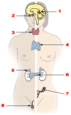
Here is a good diagram of the endocrine system. Click here. You can be given a diagram like this and you could be asked to identify the various glands.

Hypothalamus: The hypothalamus produces eight (8) hormones. Some of the hormones stimulate the pituitary gland to produce other hormones.

1. Thyrotropin-releasing hormone - This acts on the pituitary to release thyroid stimulating hormone (TSH)
2. Growth hormone-releasing hormone-This acts on the pituitary to release growth hormone (GH)
3. Gonadotropin-releasing hormone -This acts on the pituitary to release gonadotropins, namely, Follicle stimulating hormone (FSH) and Luteinizing hormone (LH).
4. Corticotropin-releasing hormone -This acts on the pituitary to release adrenocorticotropic hormone.
5. Oxytocin -This causes uterine contractions and also helps with secretion of the breast milk during lactation.
6. Dopamine - It is a neuroendocrine transmitter. Acts as a neuro-transmitter and also stimulates the release of othe hormones.
7. Vasopressin (Antidiuretic hormone -ADH) - Acts on the kidneys to prevent the lost of water via the urine.
8. Somatostatin - It inhibits the release of growth hormone.

Pituitary gland: Has an anterior (hormone-producing glandular) portion and a posterior (neural) portion which is an extension of the hypothalamus.  Two hormones ( Oxytocin and ADH) produced by the hypothalamus are stored in the posterior pituitary later release.
Four of the six pituitary hormones are tropic hormones. They regulate the function of other endocrine glands.Here are the hormones produced by the anterior portion:
1.  Growth hormone (GH): It stimulates growth of all body tissues but especially skeletal muscle and bone. GH mobilizes fats, stimulates protein synthesis, and inhibits glucose uptake and metabolism. Over production can lead to gigantism while underproduction can lead to dwarfism.
2. Thyroid-stimulating hormone (TSH): This promotes normal development and activity of the thyroid gland. 
3. Adrenocorticotropic hormone (ACTH): This stimulates the adrenal cortex to release  corticosteroids. 
4. The gonadotropins (follicle-stimulating hormone (FSH) and luteinizing hormone (LH)): These regulate the functions of the gonads in both sexes. 
     (a) FSH stimulates sex cell production.  
     (b) LH stimulates gonadal hormone production. 
5. Prolactin (PRL): This promotes milk production in humans females. 
The posterior portion stores and releases two hypothalamic hormones:
1. Oxytocin: This stimulates powerful uterine contractions during labor and delivery of babies. It also causes milk ejection in nursing women. 
2. Antidiuretic hormone (ADH): This stimulates the kidney tubules to reabsorb and conserve water. This results in the production of small volumes of highly concentrated urine and decreased plasma osmolality. Underproduction leads to a condition called diabetes insipidus, where the affected person passes a lot of diluted urine.

Thyroid gland

It produces the thyroid hormone (TH), which includes thyroxine (T4) and triiodothyronine (T3). It increases the rate of cellular metabolism. 

Calcitonin, is produced by the parafollicular cells of the thyroid gland. It decreases the blood calcium levels.

Parathyroid glands

It secretes parathyroid hormone (PTH), which causes an increase in blood calcium levels.

Pancreas

The pancreas is both an exocrine and an endocrine gland. Exocrine means that it has ducts. The endocrine portion (islets of langerhans) releases insulin and glucagon. It also releases smaller amounts of other hormones to the blood.
Glucagon, released by alpha (α) cells - It increases the glucose level in the blood.
Insulin is released by beta (β) cells - It reduces the glucose level in the blood. It increases the rate of glucose uptake and metabolism by most body cells. 

Gonads

The ovaries of the female release two main hormones - estrogens and progesterone. Estrogens stimulate maturation of the female reproductive system and development of the secondary sexual characteristics. Progesterone works with estrogens in establishing the menstrual cycle.
The testes of the male produce testosterone. It promotes maturation of the male reproductive organs, development of secondary sex characteristics, and production of sperm by the testes.

Pineal gland

The pineal gland produces the hormone  melatonin, which influences daily rhythms such as sleep and wake patterns.

Thymus 
It is an important organ of the immune system during the developmental stages of life. It vanished by the time of birth. The T-cells mature here.

METABOLIC REACTIONS

Metabolism is the building up (synthesis) or the breakdown of substances in the living organism. It consists of anabolism and catabolism.

Anabolism is the formation or synthesis of complex substances from simple ones. That is, A + B -----> C.
An example of anabolic reaction is photosynthesis.

Catabolism is the breakdown of complex substances into simple ones. That is, C -----> A + B.
An example of catabolic reaction is respiration.

What is photosynthesis?
This is the process whereby  plants produce their food (glucose) using carbon dioxide, water, and energy from the sun,  in the presence of chlorophyll (the green pigment in the leaves). Chlorophyll is contained in the chloroplast and it traps the energy from the sunlight. Glucose is the main product formed and oxygen is given off as waste.

This the equation that summarizes photosynthesis: 6CO+ 6H2à C6H12O+ 6O2
This is carbon dioxide: CO
This is water: H2O
This is glucose: C6H12O6
This is oxygen: O2

Note: You should know this equation off-hand. 

Water goes up from the roots through the plant vessel called xylem.
The glucose formed in the leaves are transported to storage sites via the plant vessel called phloem.

The glucose is stored as starch in the plants.

Respiration
Respiration involves the breakdown of glucose in the presence of oxygen to release energy in the form of ATP (ATP stands for adenosine  triphosphate, the energy currency). ATP is generated in the mitochondrium. Respiration is a combustion reaction since oxygen is needed.

There are three (3) stages in the process of respiration.
1. Glycolysis: Glucose is activated and broken down to pyruvate.
2. Kreb's cycle (also known as the Tricarboxylic Acid Cycle - TCA cycle): Some ATP, NADH, FADHare produce here.
3. Electron Transport Chain (ETC): Most ATP's are produced here.

This equation summarizes the reaction of respiration:
 C6H12O+ 6O Ã 6CO+ 6H2

Know this very well. It is simply the reverse of photosynthesis reaction.

In the absence of oxygen, pyruvate is converted into lactic acid, which  causes muscle cramps. When oxygen is available, the lactic acid is changed back to pyruvate which is then used up in the Kreb's cycle.

Fermentation
This is the process whereby glucose is changed to alcohol in the absence of oxygen.

Friday, May 2, 2014

NUCLEIC ACIDS

Today, we want to look at the structure of DNA and RNA.

STRUCTURE OF DNA
DNA stands for deoxyribonucleic acid. This is the basic unit of our genes. DNA consist of a sugar molecule called deoxyribose, a phosphate, and nitrogen bases. These three things make up the nucleotide. So we can say that the basic unit of the DNA is the nucleotide. The sugar and the phosphate together make up the nucleoside.

The nitrogen bases are adenine (A), guanine (G), cytosine (C), and thiamine (T).
Adenine (A) always binds to thiamine (T)
Guanine (G) always binds to cytosine (C)

DNA is located in the nucleus of the cell and it has a double helix structure.

STRUCTURE OF RNA
RNA stands for ribonucleic acid. This is involved with the synthesis of protein. RNA consist of a sugar molecule called ribose, a phosphate, and nitrogen bases. These three things make up the nucleotide. So we can say that the basic unit of the RNA is the nucleotide. The sugar and the phosphate together make up the nucleoside.

The nitrogen bases are adenine (A), guanine (G), cytosine (C), and Uracil (U).
Adenine (A) always binds to Uracil (U)
 Guanine (G) always binds to cytosine (C)

RNA is located in the cytoplasm of the cell and it is a single strand.

Types of RNA:
1. mRNA--- Messenger RNA: This is code for protein synthesis. 
2. tRNA ---- Tranfer RNA: This carries the amino acids.
3. rRNA ---- Ribosomal RNA: This reads the code on the mRNA.

Note the difference between DNA and RNA. DNA has thiamine but no uracil. RNA has uracil but no thiamine.